


Explore Any Narratives
Discover and contribute to detailed historical accounts and cultural stories. Share your knowledge and engage with enthusiasts worldwide.
For over a century, scientists have known the influenza virus as a shape-shifting enemy. We’ve mapped its genetic code, tracked its global spread, and developed vaccines that chase its ever-mutating form. Yet a fundamental mystery remained shrouded in darkness: the precise, nanoscopic moment a single virus particle tricks a human cell into letting it inside. All we had were blurry snapshots and educated guesses. That changed in December 2025.
A collaboration between Swiss and Japanese scientists published a study that didn't just add another chapter to virology—it rewrote the opening scene. Using a novel hybrid microscope they built from the ground up, the team achieved the first real-time, high-resolution visualization of an influenza virus invading a living human lung cell. What they witnessed overturned a foundational assumption. The cell isn’t a passive victim. It’s an active, if misguided, participant. Infection, it turns out, is a tragic pas de deux performed on a stage one ten-thousandth the width of a human hair.
The technical achievement here is staggering. Previous methods forced a brutal choice. Electron microscopy could show exquisite structural detail, but only by flash-freezing and slicing the cell, murdering it for a single post-mortem photograph. Fluorescence microscopy could watch living cells, but its resolution was too poor to see the virus itself; you’d see only a blurry dot of light. The new technique, dubbed ViViD-AFM (Virus-view dual confocal and Atomic Force Microscopy), smashes that compromise. It combines the precise, physical probe of an atomic force microscope—which feels the surface of the cell like a blind person reading braille—with the dynamic color tracking of a confocal laser. The result is a live-action film where every actor, from viral spike to cellular protein, has a defined role.
"We are no longer inferring the steps of entry from before-and-after pictures. We are watching them unfold, in real time, with our own eyes. The narrative of viral infection is now a documentary," said Dr. Petra Müller, a biophysicist at the University of Basel who co-led the work, in an interview accompanying the publication.
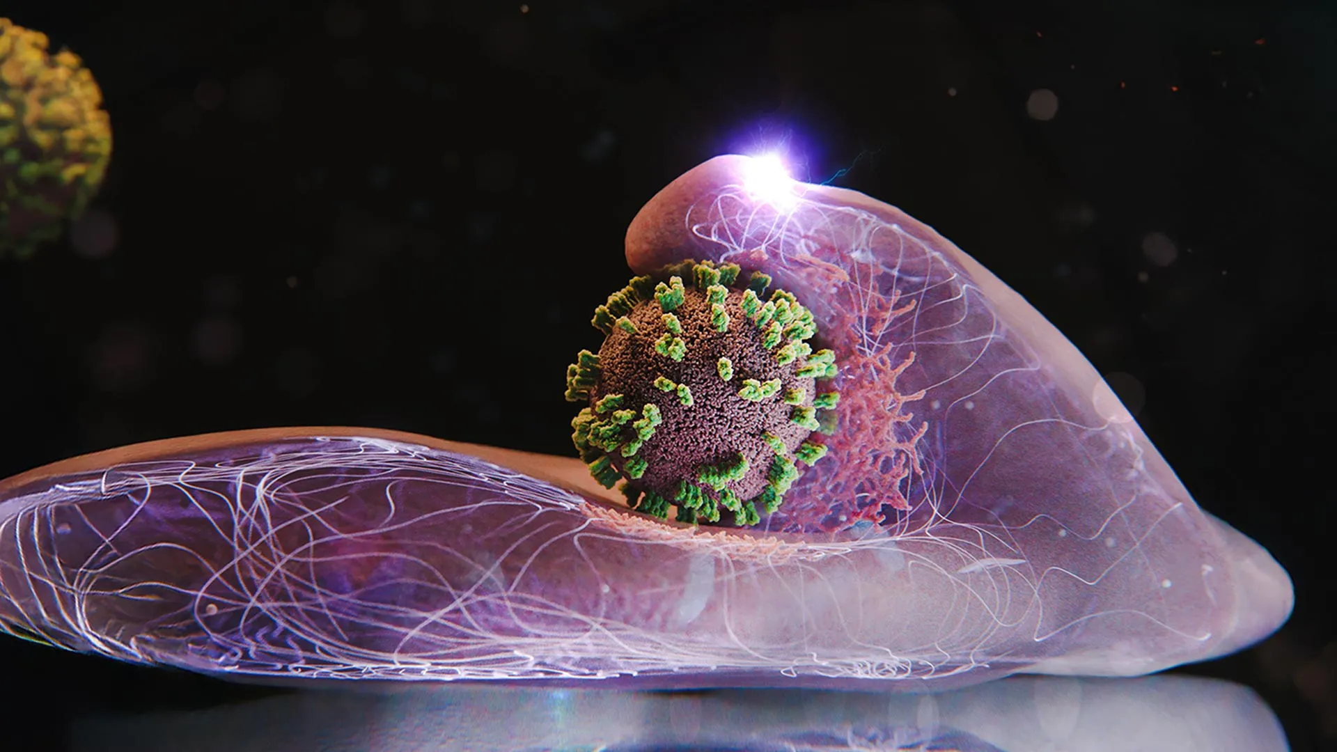
The traditional textbook illustration is simple: a spiky virus ball bumps into a cell, fuses with the membrane, and empties its genetic cargo. Neat. Passive. Wrong. The ViViD-AFM footage reveals a far more dynamic and unsettling sequence. When an influenza virion makes contact, the cell membrane doesn’t just sit there. It responds. It bulges upward, forming a kind of molecular hill beneath the virus. If the virus starts to drift away, these membrane undulations intensify, as if the cell is actively trying to corral it back.
This is the first critical discovery: the cell’s own machinery is hijacked from the very first touch. The researchers watched as clouds of clathrin, a protein the cell uses to import nutrients, rushed to the site of viral contact. Clathrin’s normal job is to form a little coated pit that buds inward, bringing essential molecules into the cell. Influenza has evolved to issue a forged work order. The cell obediently starts constructing a clathrin-coated pit around the virus, essentially building the Trojan horse that will carry its enemy past the gates.
Yohei Yamauchi, a virologist at ETH Zurich and senior author on the study, framed it with elegant simplicity. "The infection of our body cells is like a dance between virus and cell," he said. "But it’s a dance where the cell is following the virus’s lead, believing it’s waltzing with something else entirely."
"Seeing the membrane actively rise to meet the virus was the 'eureka' moment. It completely flips the script. We’re not looking at a break-in. We’re looking at a deception so perfect the guard holds the door open," Yamauchi stated.
While the Swiss-Japanese team was filming the live drama, another group on the other side of the world was creating ultra-high-definition 3D models of the key scene. At the University of Washington’s Lee Lab, researchers used cryo-electron microscopy (cryo-EM) to capture the influenza virus in the very act of membrane fusion. Their work, published in the Journal of Virology in late 2025, provides the structural playbook for the dance.
Cryo-EM works by firing electrons through a virus sample frozen in a thin layer of ice at -196° Celsius. This preserves the native structure of proteins in mid-action. The Lee Lab’s images reveal, at the atomic level, how the virus’s hemagglutinin "spike" protein undergoes a catastrophic rearrangement. It’s a molecular jack-in-the-box. Upon sensing the acidic environment inside the cellular pit, the spike protein refolds violently, stabbing a fusion peptide into the host membrane and zipping the two membranes together. This creates the channel through which the viral genome floods into the cell’s cytoplasm.
The synergy between these two studies is powerful. ViViD-AFM shows the *when* and *where* of the cellular response in living color. Cryo-EM reveals the *how* of the viral machinery in atomic detail. Together, they form the most complete picture of influenza entry ever assembled. The Lee Lab’s structural data shows the virus’s precise tool for forcing fusion; the ViViD-AFM footage shows the cell naively providing the ideal workspace for that tool to operate.
One minor but fascinating detail from the cryo-EM work involves the lipid composition of the membranes themselves. The fusion doesn’t happen just anywhere. The virus appears to seek out, or induce, specific lipid microdomains on the cell surface—slightly more disordered, slightly more flexible patches of membrane that are easier to merge with. It’s a burglar checking for the loosest window latch.
The immediate application is visceral. For the first time, pharmaceutical researchers can watch an antiviral drug *work*—or fail—in real time. Instead of waiting 48 hours to count dead cells in a plate, they can add a candidate compound to a culture and use ViViD-AFM to see if it blocks the membrane bulging, disrupts the clathrin recruitment, or paralyzes the viral fusion spike. This turns drug screening from a slow, inferential process into a direct observational science.
Imagine testing a new flu drug. The old way: apply the drug, infect the cells, wait a day or two, stain the cells, and measure the signal. You get a number—a percentage of inhibition. The new way: apply the drug, introduce the virus, and watch. Does the virus still get grabbed? Does the clathrin still swarm? You get a *movie* of failure, a direct visual of the mechanism. This accelerates the pipeline and, more importantly, provides immediate insight into *why* a drug works. Is it blocking attachment? Arresting fusion? That knowledge is gold for designing the next generation of therapeutics.
The implications spill far beyond influenza. The ViViD-AFM technique is a platform. HIV, herpes, dengue, Zika, SARS-CoV-2—any enveloped virus that enters cells via membrane fusion is now a candidate for this kind of live-cell interrogation. Furthermore, the method isn't limited to pathogens. The same principles can track how lipid nanoparticles in mRNA vaccines deliver their cargo, or how targeted cancer drugs are internalized by tumor cells. We have been handed a universal microscope for viewing the intricate, invisible handshake between any particle and a living cell.
This is not incremental progress. It is a paradigm shift in cellular microbiology. For decades, we’ve been piecing together a crime scene from static evidence. Now, we have the security camera footage. And the first viewing reveals we misunderstood the crime entirely. The butler didn’t just leave the door unlocked. He escorted the thief inside, hung up his coat, and pointed him toward the silver.
The influenza virus, a seemingly simple lipid sphere studded with proteins, has long been underestimated in its cunning. What the new ViViD-AFM system, a marvel of bioengineering from Switzerland and Japan, reveals is not just how the virus enters, but how the cell, in its complex biological naiveté, facilitates its own demise. This hybrid microscopy, combining the tactile precision of atomic force microscopy with the visual clarity of confocal fluorescence, allows scientists to "follow the detailed dynamics of the virus's entry into the cell" in real time, a capability previously confined to science fiction.
Professor Yohei Yamauchi, a leading figure in molecular medicine at ETH Zurich, spearheaded this groundbreaking effort. His team’s work, published in Cell Discovery in December 2025, fundamentally reshapes our understanding of viral pathogenesis. For years, electron microscopy offered only frozen, lifeless snapshots, forcing researchers to infer dynamic processes from static images. Fluorescence microscopy, while allowing live observation, lacked the spatial resolution to discern the fine details of viral interaction. ViViD-AFM eradicates these limitations, providing an unprecedented, high-definition, live-action view of the cellular invasion.
"We had theories, elegant models, but seeing is believing. The cell's active participation was not just unexpected; it was a profound re-evaluation of the host-pathogen relationship," explained Professor Yamauchi in a press conference following the publication.
The most startling revelation from the ViViD-AFM footage is the active role played by the host cell. Far from being a passive barrier, the cell engages in a series of molecular maneuvers that directly aid the virus. Upon contact, the cell surface doesn't merely deform; it actively bulges upward, almost enveloping the viral particle. These wave-like membrane movements intensify if the virus attempts to detach, as if the cell is exerting a subtle, magnetic pull to re-engage its attacker. This is not a struggle; it is a misplaced embrace.
The machinery behind this tragic embrace involves actin, a protein crucial for maintaining cell shape and movement. The bulges observed are composed of actin, indicating a deliberate, energy-intensive cellular response. Simultaneously, the cell actively recruits clathrin proteins to the site of viral attachment. Clathrin’s normal function is to form coated pits for internalizing essential substances like hormones, iron, and cholesterol. The influenza virus, a master manipulator, exploits this fundamental cellular pathway. It’s a classic Trojan horse scenario, but with the added twist that the city's guards are actively building the horse and pulling it through the gates themselves.
This hijacking of normal cellular machinery represents a critical vulnerability. The virus isn't inventing new pathways; it's simply diverting existing ones for its own nefarious ends. The process culminates in the virus being "pinched off into a vesicle and carried deeper into the cell, toward the nucleus," a journey that ultimately leads to viral replication and the infection of neighboring cells. Is the cell truly helpless, or is there a molecular signal, a receptor, that could be blocked to prevent this fatal hospitality?
"The ongoing surge in flu cases across major metropolitan areas like New York City, with emergency rooms overflowing in December 2025, underscores the urgent need for new insights into viral entry. This microscopy technique is not just academic; it has immediate, tangible implications for public health," stated Dr. Lena Hansen, an epidemiologist at Columbia University, speaking on the critical relevance of the research.
While the initial focus of ViViD-AFM has been influenza, its potential applications stretch across the entire spectrum of virology and cellular biology. The system’s ability to distinguish between different influenza subtypes based on their physical behavior—demonstrating that H1N1 and H3N2, for example, exhibit statistically different diffusion coefficients—is profoundly significant. This means researchers can rapidly assess how minor mutations in viral surface proteins might alter entry dynamics, providing an invaluable tool for evaluating emerging variants and predicting their infectivity.
Furthermore, the technique’s sensitivity is remarkable. Influenza exhibits relatively weak interactions with cell membranes, with binding forces measuring a mere 10-25 piconewtons. Despite these delicate forces, ViViD-AFM successfully visualized the entire entry process. This suggests the system can be applied to virtually any virus, including those with stronger receptor affinities, and even to other non-viral cellular interactions. Imaging the entry of HIV, herpes, dengue, or Zika viruses now seems within reach, promising a cascade of new discoveries.
"The new technique therefore provides key insights when it comes to the development of antiviral drugs and is suitable for testing the efficacy of potential drugs in a cell culture in real time," asserted Professor Yamauchi, emphasizing the immediate practical utility of the discovery for pharmaceutical research.
This real-time drug testing capability is a game-changer. Instead of relying on indirect assays that measure viral replication after the fact, scientists can directly observe if a candidate drug prevents the initial cellular capture, inhibits the clathrin recruitment, or blocks the membrane fusion. This accelerates the drug discovery pipeline dramatically, allowing for rapid iteration and refinement of compounds. The days of blind screening, hoping for an effect without understanding the mechanism, are slowly drawing to a close. We can now pinpoint precisely where a drug interrupts the viral life cycle, enabling the development of more targeted, and therefore more effective, therapies.
The applications extend even further. Imagine tracking how lipid nanoparticles, the delivery vehicles for modern mRNA vaccines, interact with cells. Or observing how drug-carrying nanoparticles, designed for targeted cancer therapies, enter tumor cells. The ViViD-AFM system is not just a flu microscope; it is a universal platform for understanding the nanoscopic ballet between cells and the particles that interact with them, whether those particles are friend or foe. Its emergence in late 2025, amidst an ongoing public health crisis, was a stark reminder of the relentless pursuit of scientific understanding in the face of biological threats.
The true weight of the December 2025 findings from the Yamauchi lab and the University of Washington extends far beyond a single virus. This work represents a fundamental shift from inferential biology to observational truth. For over a century, virologists have been like detectives reconstructing a bank heist from a smashed vault door and scattered money. They knew the outcome and had clues to the method, but they never saw the robbers in action. ViViD-AFM and complementary cryo-EM have installed a 24/7 surveillance system inside the bank. The significance is not just in catching one criminal, but in understanding the blueprint of the crime itself—a blueprint that many pathogens share.
This changes how we teach microbiology. The next generation of textbooks will need new illustrations. The static image of a virus bumping into a cell will be replaced by a multi-panel sequence showing the active membrane bulging, the clathrin recruitment, and the vesicular pinching. It reframes our entire immunological perspective. Our cells are not inert fortresses; they are dynamic, responsive entities that can be tragically fooled. This has profound implications for understanding autoimmune diseases, cancer metastasis, and even neurological conditions where cellular communication goes awry. The technique provides a universal lens for observing any interaction at the cellular surface.
"This is akin to the invention of the telescope for astronomers. We are not just seeing farther; we are seeing differently. The cell is not a static bag of chemicals. It is a responsive, interactive entity, and we now have a front-row seat to its conversations—even the disastrous ones," stated Dr. Aris Kotsis, a cellular biophysicist at the Weizmann Institute of Science, commenting on the broader implications.
For all its revolutionary power, this new microscopy is not a panacea, and treating it as such would be a scientific misstep. The first and most obvious limitation is throughput. The ViViD-AFM system is painstakingly complex, requiring a perfectly tuned environment and expert operation. It cannot yet scan thousands of cells simultaneously like a high-throughput screening assay. This means its initial role in drug discovery will likely be as a deep-dive validation tool for promising candidates identified by faster, cruder methods, not as a primary screen for millions of compounds.
A more subtle, but critical, limitation involves the artificiality of the cell culture environment. The team’s stunning footage was captured using immortalized human lung epithelial cells growing in a petri dish. This is a clean, simplified model. It lacks the chaotic, three-dimensional reality of actual lung tissue, complete with mucus, cilia, immune cell patrols, and the complex extracellular matrix. Does the virus employ the exact same "dance" when faced with the multicellular architecture and immune pressures of a living organism? The answer is probably not entirely. The observed clathrin-mediated entry might be one pathway among several, its importance shifting based on tissue type and host defenses.
Furthermore, the technique’s intense focus on the very first moments of contact risks creating a myopic view of infection. Viral success is not determined solely by entry efficiency. Replication, assembly, immune evasion, and transmission are equally critical stages. A drug that perfectly blocks the entry dance observed by ViViD-AFM might simply select for viral mutants that use an alternate, unseen back door. The real-world efficacy of therapeutics developed from these insights remains to be proven in clinical trials, a path that remains long, expensive, and fraught with failure. The microscope shows us a potential point of failure, but biology has a notorious habit of finding workarounds.
The immediate trajectory for this research is clear and concrete. The Yamauchi lab has already announced plans to apply ViViD-AFM to SARS-CoV-2 variants, with initial comparative studies scheduled for publication in the third quarter of 2026. Parallel work is underway at the Lee Lab to solve the cryo-EM structures of influenza fusion intermediates bound to novel inhibitory antibodies, work funded by a new NIH grant and expected to yield pre-print data by late 2026. These are not vague aspirations; they are the next scheduled experiments on the lab calendars.
The pharmaceutical industry is watching closely. Several major drugmakers, including Gilead and Roche, have initiated collaborative agreements to use adapted versions of this imaging technology within their own antiviral discovery pipelines. The goal is not just for flu. The real prize is a universal, observation-driven platform to combat the next pandemic virus—the one we haven't met yet. By understanding the shared choreography of viral entry, we can design broader-spectrum entry inhibitors, moving from virus-specific drugs to pathogen-class therapeutics.
On a longer timeline, the technology itself will evolve. The current iteration of ViViD-AFM is a bespoke instrument in a specialized lab. The next five years will see a push for commercialization and miniaturization, making the technology more accessible to academic and clinical labs worldwide. The dream is a standardized "virological observatory" that can be deployed during an outbreak to rapidly characterize how a novel pathogen breaches cells, informing public health responses in real time.
The dance of infection, once a theoretical abstraction, is now a visible, measurable phenomenon. We have seen the steps. The question is no longer how the virus leads, but how we can cut in. The final move belongs to us.
Your personal space to curate, organize, and share knowledge with the world.
Discover and contribute to detailed historical accounts and cultural stories. Share your knowledge and engage with enthusiasts worldwide.
Connect with others who share your interests. Create and participate in themed boards about any topic you have in mind.
Contribute your knowledge and insights. Create engaging content and participate in meaningful discussions across multiple languages.
Already have an account? Sign in here

MIT chemists synthesize verticillin A after 55 years, unlocking a potential weapon against fatal pediatric brain tumors ...
View Board
Radiation-driven wolves in Chernobyl display rapid cancer-resistant evolution, a 30-year natural experiment revealing ge...
View Board
Cancer research reaches new heights as ISS microgravity enables breakthroughs like FDA-approved pembrolizumab injections...
View Board
Pancreatic cancer's sugar-coated shield uncovered: Researchers reveal how tumors exploit sialic acid to deceive immune c...
View Board
CERN's Large Hadron Collider achieves alchemy, converting lead to gold via ultra-peripheral collisions, validating quant...
View Board
Descubre cómo Carl Wieman, premio Nobel de Física, revolucionó la educación científica. De atrapar átomos a transformar ...
View Board
Entdecken Sie das Leben von Robin Warren, dem medizinischen Pionier, der mit der Entdeckung von Helicobacter pylori die ...
View Board
2026 marks a pivotal year for mRNA tech, with breakthroughs in cancer, HIV, microneedles, and AI-driven trials set to re...
View Board
MIT researchers transform ordinary concrete into structural supercapacitors, storing 10x more energy in foundations, tur...
View Board
Microbial oceanographer Dr. Anya Sharma's 2025 discovery of deep-sea bacteria actively fixing carbon challenges decades ...
View Board
Scientists reverse blood stem cell aging with lysosomal inhibitors and RhoA blockers, restoring regenerative capacity an...
View Board
Kathmandu’s 1,324‑meter high valley blends 1596 Kasthamandap, UNESCO temples and Newar brickwork with four million resid...
View Board
MIT’s 2026 breakthroughs reveal a world reshaped by AI hearts, gene-edited embryos, and nuclear-powered data centers, wh...
View Board
Un rein de porc génétiquement modifié par CRISPR a sauvé un patient en 2024, marquant un tournant historique dans la lut...
View Board
Le loup terrible, éteint depuis 10 000 ans, renaît dans un labo texan : trois louveteaux génétiquement modifiés, Romulus...
View Board
The Strange Connection Between Brutalist Architecture and Soviet Propaganda A concrete postcard from 1971 shows a famil...
View Board
Journalist details Walter Freeman's 1946 ice-pick lobotomy, its 10-minute office speed, 2,500 operations, and the enduri...
View Board
The 1174 Canterbury Cathedral Fire: An Architectural Phoenix Smoke, thick and acrid, choked the Kentish sky on the fift...
View Board
Major 2025 trials reveal no effective treatments for long COVID brain fog, forcing a shift from cognitive training to im...
View Board
Spaceflight rapidly rewrites human gene expression, accelerating aging and stressing stem cells, as new studies reveal p...
View Board
Comments